|
Human Embryo at Blastocyst Stage View 1
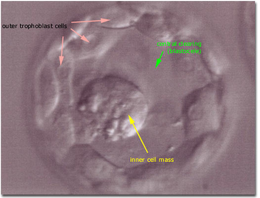
Photomicrograph (microscopic view) of two different human blastocysts, which form after the solid mass of blastomeres that comprise a morula undergo rearrangements to form an embryo with a central area of clearing (the blastocele), an inner cell mass (projecting into the blastocele), and a thin outer layer of trophoblast cells (that line the blastocele). Blastocysts are generally seen in the In Vitro Fertilization laboratory after 120 hours (5 days) from the time of insemination (on post- egg retrieval day 5).
|

|