|
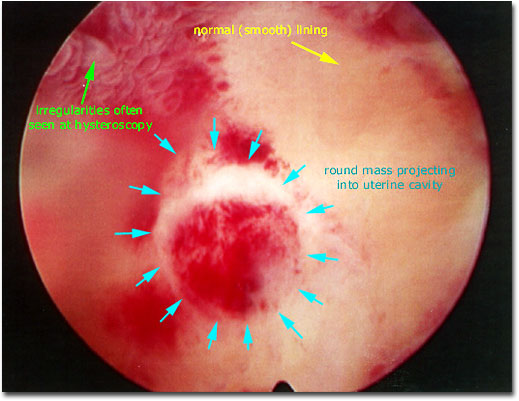
Photograph of a hysteroscopic view of the uterine cavity identifying a round mass that is projecting into the cavity. This mass could either be an endometrial polyp (composed of endometrium that is organized into a polypoid structure that usually has its own prominent vasculature = blood vessels) or a submucosal leiomyoma (fibroid composed of smooth muscle cells derived from cells of the uterine wall and projecting into the cavity).
Surgical resection with a resectoscope is straightforward when the stalk of the mass can be identified (as in this case). Electrocautery is rarely required if this is an endometrial polyp but can be readily applied if the mass is composed of muscle (a denser type of tissue than endometrium). In this case, the mass was easily removed without the use of electrocautery and a pathologist identified a benign endometrial polyp using microscopy.
|

|