|
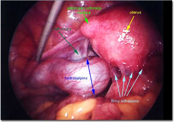
Photograph of a pelvis that demonstrates the uterus (densely adherent to surrounding structures), left fallopian tube (markedly dilated and adherent to surrounding structures), and bowel (densely adherent to the uterus and pelvic organs). Note the rounded distended section of uterine tissue at the interface of the left fallopian tube and uterus, usually found to be salpingitis isthmica nodosa (thought to be a result of chronic local inflammation in which the lumen of the isthmic section of the fallopian tube contains diverticulae into the surrounding muscular wall of the tube).
In order to repair such a fallopian tube, the pelvic structures must (ideally) be completely freed from one another. These adhesions are predominantly filmy yet they are extensive. These adhesions are often the result of a prior local infection so prophylactic antibiotic therapy is advisable.
|

|